
What Causes Varicose Veins and When to See a Specialist
Healthy blood circulation is a cornerstone of overall physical well-being, yet circulatory issues in the lower extremities are frequently misunderstood or overlooked. Millions of people worldwide notice bulging, twisted vessels on their legs, often dismissing them as an unavoidable aesthetic consequence of aging. However, these visible changes are frequently the external signs of an underlying medical condition known as chronic venous insufficiency (CVI). Understanding what causes varicose veins, identifying the early warning signs of venous disease, and knowing when to seek professional medical evaluation are crucial steps in protecting long-term vascular health.
Understanding Varicose Veins and Blood Flow
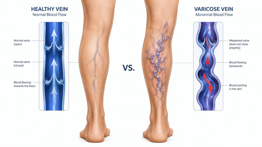
To comprehend why varicose veins develop, it is necessary to examine how the circulatory system operates against gravity. The vascular network relies on a complex mechanism to return oxygen-depleted blood from the lower extremities back to the heart. While arteries carry oxygen-rich blood away from the heart under high pressure, veins must transport blood upward, working directly against gravitational pull. [Normal Vein] --> Valves close properly --> Blood flows upward toward the heart [Diseased Vein] --> Valves fail to close --> Blood flows backward (Reflux) & pools To prevent blood from flowing backward, veins are equipped with tiny, one-way bivalve leaflets called vein valves. When the calf and leg muscles contract during movement, they act as a natural pump, squeezing the veins and pushing blood upward. As the muscle relaxes, the one-way valves snap shut, anchoring the blood in place and preventing it from slipping back down toward the feet. When these delicate valves become stretched, weakened, or structurally damaged, they lose their ability to close completely. This mechanical failure allows blood to flow backward and pool within the lower segments of the vessel—a pathological process termed venous reflux. Over time, the continuous accumulation of pooled blood increases the internal pressure within the vein. This chronic hydrostatic pressure forces the vessel walls to stretch, widen, and twist, eventually causing them to protrude beneath the surface of the skin as swollen veins.
What Causes Varicose Veins?
The root cause of varicose veins is vein valve dysfunction, but the structural degradation of the vein wall itself is equally significant. Healthy vein walls possess natural elasticity, allowing them to expand and contract to accommodate fluctuations in blood volume. If the collagen and elastin fibers within the venous walls degrade, the vessel loses its structural integrity. As the vein wall stretches permanently, the valve flaps are pulled apart, preventing them from meeting in the center to seal the vessel. This creates a cyclical, self-perpetuating problem: wall weakness exacerbates valve failure, and the resulting pooling of blood further stretches the weakened vein wall. While superficial spider veins the tiny, web-like clusters of red, blue, or purple vessels near the skin’s surface are primarily cosmetic concerns stemming from micro-circulatory changes, varicose veins indicate a deeper structural failure within the larger superficial venous system, such as the great saphenous vein.
Risk Factors That Increase Vein Problems
While anyone can develop venous reflux, several distinct clinical and constitutional risk factors significantly elevate the likelihood of developing blood circulation disorders. Genetic Predisposition: Heredity is the single most dominant risk factor for venous insufficiency. If both parents have a history of varicose veins, an individual’s statistical likelihood of developing the condition exceeds 80%. This genetic link typically involves inherited structural deficiencies in the strength of the vein walls or the formation of the venous valves. Biological Sex and Hormonal Fluctuations: Females are diagnosed with varicose veins at a significantly higher rate than males. This disparity is primarily driven by hormonal influences. Progesterone and estrogen, which fluctuate during menstrual cycles, pregnancy, and menopause, naturally relax the smooth muscle tissue within vein walls, making the vessels more susceptible to stretching and valve separation. Advanced Age: The natural aging process causes a gradual loss of muscle mass and skin elasticity, alongside the wear and tear of the delicate valve leaflets inside the veins. Over decades of continuous gravitational pressure, these valves become inherently less efficient.
Lifestyle and Occupational Contributors
Beyond unmodifiable biological risk factors, daily habits and occupational demands play a decisive role in the acceleration of chronic venous insufficiency.
ContributorPathological Impact on CirculationProlonged StandingContinuous hydrostatic pressure forces blood downward without the assistance of calf muscle contractions, accelerating valve failure.Sedentary SittingThe calf muscle pump remains completely inactive, causing blood to pool in the deep and superficial veins of the lower legs.Obesity & WeightExcess body mass increases intra-abdominal pressure, restricting the smooth upward return of blood from the legs to the torso.Lack of ExerciseWeakened lower extremity muscles reduce the efficiency of the mechanical pumping required to clear blood from the lower limbs.
Early Symptoms People Often Ignore
Venous disease is notoriously progressive, frequently starting with subtle, non-visual sensations that individuals mistakenly attribute to general fatigue or a busy lifestyle. Recognizing these early signs of vein disease symptoms is critical for timely intervention before permanent vascular changes occur.
- Persistent Leg Fatigue and Heaviness: A classic early indicator is a profound sensation of heavy legs, which typically worsens as the day progresses, particularly in the late afternoon or evening.
- Mild, Deep-Seated Aching: Patients often describe a dull, generalized ache throughout the calves or thighs after periods of inactivity.
- Localized Burning and Itching: As blood pools, it can trigger localized inflammatory responses in the surrounding tissues, causing a persistent burning sensation or intense itching directly over or near the affected superficial vessels.
Signs That Vein Disease May Be Progressing
When early circulation problems are left unaddressed, the structural damage within the venous network deepens, moving from a mild annoyance to a more restrictive medical condition.
Leg Pain and Swelling
As venous reflux intensifies, fluid begins to leak out of the high-pressure veins and filter into the surrounding soft tissues of the lower leg, ankles, and feet. This fluid retention, known medically as venous edema, causes visible leg pain and swelling. Individuals may notice that their socks leave deep, lasting indentations around their ankles at the end of the day, or that their shoes feel uncomfortably tight.
Nighttime Leg Cramps and Restlessness
The accumulation of metabolic waste products in the stagnant, pooled venous blood can irritate localized nerves and muscle fibers. This frequently manifests as painful nighttime leg cramps, commonly referred to as "charley horses." Additionally, many patients experience an irresistible urge to move their legs during periods of rest a presentation closely linked to secondary Restless Legs Syndrome (RLS) driven by vascular congestion.
Skin Changes and Advanced Vein Symptoms
If chronic venous insufficiency remains untreated over several years, the high pressure within the venous system causes red blood cells to break down and leak into the surrounding skin tissue. This progression marks the transition into advanced stages of vascular disease. Chronic Venous Insufficiency ↳ Red Blood Cells Leak into Tissue ↳ Hemosiderin Deposition (Rust-Colored Staining) ↳ Lip dermatosclerosis (Skin Hardening & Inflammation) ↳ Venous Stasis Ulcers (Open, Non-Healing Wounds) The breakdown of red blood cells releases iron into the skin, leading to a permanent, rust-colored brownish discoloration known as hemosiderin staining. The skin over the lower third of the leg can become chronically inflamed, dry, leathery, and hardened a condition termed lip dermatosclerosis. In its most severe presentation, the compromised skin loses its nutrient supply entirely, breaking down to form painful, open, non-healing sores called venous stasis ulcers. These ulcers typically form around the inner ankle bones and are highly susceptible to recurrent bacterial infections.
When to See a Specialist for Varicose Veins
It is a common misconception that a person should only schedule a vein consultation if their veins are visually prominent or causing severe, debilitating pain. Seeking an evaluation from a qualified vascular professional early can prevent the painful and complex complications associated with advanced disease progression.
You should seek a formal medical evaluation if you experience any of the following indicators:
- Leg pain, throbbing, or swelling that does not improve after elevating your feet.
- Skin alterations, including unexplained hyperpigmentation, redness, or textural hardening around the calves or ankles.
- The appearance of open sores, breaks in the skin, or weeping fluid in the lower legs.
- Spontaneous bleeding from a varicose vein, which constitutes a medical emergency due to the high pressure within the vessel.
- A sudden increase in localized swelling, warmth, or tenderness, which may indicate a deep vein thrombosis (DVT) or superficial thrombophlebitis (blood clots).
For those navigating these symptoms, consulting a board-certified vascular specialist ensures an accurate diagnosis. Highly regarded experts in the field, such as Dr. Amir Salem, MD, emphasize that addressing venous insufficiency in its earlier stages significantly reduces the long-term risk of skin damage and irreversible tissue changes.
How Vein Specialists Diagnose Vein Disease
A modern vascular evaluation is entirely non-invasive, painless, and completed in an outpatient setting. The diagnostic process begins with a comprehensive review of the patient’s medical history, lifestyle factors, and an objective physical examination of the lower limbs while standing.
The Diagnostic Gold Standard: The core tool used by a vascular doctor to evaluate circulation is an ultrasound vein scan, specifically a duplex ultrasound evaluation.
This specialized diagnostic imaging tool combines traditional ultrasound formatting with Doppler technology to accomplish two vital goals:
- Anatomical Mapping: It provides a clear, real-time visual image of both the deep and superficial venous structures, mapping out the precise location of any structurally abnormal vessels.
- Hemodynamic Assessment: It measures the exact speed and direction of blood flow through the veins. This allows the specialist to pinpoint precisely which vein valves are failing and quantify the exact duration of the venous reflux.
Modern Treatment Approaches for Varicose Veins
The historical era of invasive, painful vein stripping surgeries requiring hospital stays and prolonged recovery periods has been entirely replaced by modern, minimally invasive vein treatment options. Today's procedures are performed in an office setting under local anesthesia, allowing patients to walk out of the clinic and return to normal activities almost immediately.
- Endo venous Laser Ablation (EVLA): A highly precise procedure where a specialist inserts a microscopic laser fiber directly into the diseased vein through a tiny puncture site. The laser delivers targeted thermal energy to the interior walls of the vessel, causing the malfunctioning vein to safely collapse and seal shut.
- Radiofrequency Ablation (RFA): Similar to EVLA, this technique utilizes controlled radiofrequency energy instead of laser light to heat, contract, and permanently close the targeted insufficient vein.
- Sclerotherapy: This approach is primarily used for smaller varicose veins and spider veins. A specialized medicinal solution or foam is injected directly into the target vessel. The solution irritates the lining of the vein, causing it to stick together and clot off safely. Over time, the body naturally absorbs the closed vessel tissue.
- Non-Thermal Vein Closure (Adhesives/Mechanochemical): Advanced treatments like Vena Seal utilize a medical-grade adhesive inserted via a tiny catheter to seal the diseased vein instantly without the need for heat or multiple numbing injections.
Once the diseased, non-functioning vein is successfully closed by a varicose vein specialist, the body automatically reroutes the pooled blood into nearby, healthy, functional veins. This immediately restores efficient, unhindered blood flow back to the heart, resolving tissue congestion and eliminating painful symptoms.
Risks of Ignoring Vein Symptoms
Neglecting symptoms of venous reflux allows the underlying circulatory failure to worsen unchecked. Beyond cosmetic changes, permanent physiological consequences can arise over time. The persistent pooling of sluggish blood significantly increases the risk of superficial thrombophlebitis, which involves painful blood clots and inflammation in the veins near the skin's surface. While these superficial clots are distinct from the life-threatening clots found in deep vein thrombosis, they cause significant localized pain and can propagate into the deep venous system if unmanaged. Furthermore, the chronic tissue congestion caused by delayed treatment impairs the local immune response in the lower limbs, making the skin highly vulnerable to cellulitis a rapidly spreading, dangerous bacterial skin infection.
Conclusion
Varicose veins are not a superficial flaw or a minor inconvenience; they are a visible indicator of an underlying mechanical failure within the circulatory system. Understanding how vein valve dysfunction leads to chronic venous insufficiency empowers individuals to recognize early warning signs before severe skin damage or ulceration develops. With modern diagnostics like duplex ultrasound imaging and highly effective, minimally invasive treatments, managing vein disease is safer and more accessible than ever before. Prioritizing an early evaluation with a qualified professional can protect your long-term mobility, eliminate daily discomfort, and preserve your overall vascular health for years to come.
Frequently Asked Questions (FAQs)
What is the primary difference between spider veins and varicose veins?
Spider veins are small, thin, web-like vessels (red, blue, or purple) located just beneath the surface of the skin that do not bulge or cause structural tissue swelling; they are typically cosmetic. Varicose veins are larger, thick, bulging, and twisted vessels that indicate structural failure of deeper venous valves and underlying chronic venous insufficiency.
Can lifestyle changes alone completely cure varicose veins?
No, lifestyle modifications cannot reverse or repair a vein valve that has already suffered mechanical failure. Wearing graduated compression stockings, elevating the feet, exercising, and maintaining a healthy weight can significantly manage symptoms and prevent new varicose veins from forming, but they cannot restore a permanently damaged vein structure.
Is an ultrasound vein scan painful or invasive?
No, a diagnostic duplex ultrasound vein scan is completely non-invasive and painless. It utilizes sound waves to create visual images of your veins and evaluate blood flow dynamics. The technician simply applies a clear gel and moves a smooth wand (transducer) over the surface of your legs.
How long is the recovery period after a minimally invasive vein treatment?
Recovery from modern treatments like endo venous laser ablation or sclerotherapy is remarkably brief. Most patients walk out of the clinic immediately following the procedure and return to desk jobs or light daily activities the exact same day. Heavy lifting and high-impact workouts are generally avoided for only a few days to a week.
What happens to the blood flow once a varicose vein is closed or removed?
When a malfunctioning vein is medically closed, your body’s circulatory network automatically adapts. The blood is naturally and instantly rerouted through neighboring, healthy veins that possess properly functioning valves, which actually improves your overall leg circulation.


Velki Login: দ্রুত অ্যাকাউন্ট অ্যাক্সেসের সহজ নিয়ম
Velki Login: দ্রুত অ্যাকাউন্ট অ্যাক্সেসের সহজ নিয়ম